Digital Histology Facility
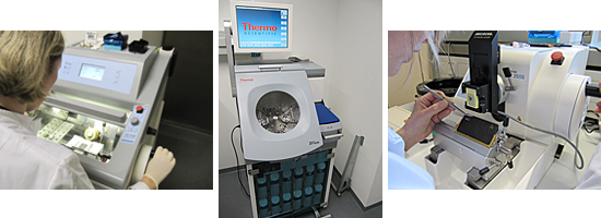
The TIGA's Digital Histology Facility provides access to the following services:

Wet lab services
- Tissue processing
- Tissue sectioning (Cryo and FFPE samples)
- Tissue staining (brightfield, fluorescence)
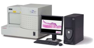
Scanning services
- Scanning of brightfield or fluorescent samples
- Digitization in 20x or 40x magnification
Image
analysis services
Depending on the
complexity of the projects, we use software frameworks to perform the following
image analysis tasks:

TMA (Tissue-microarray) quantification:
- TMAs stained in brightfield
-
TMAs
stained in fluorescence
Complete Cell Quantification
Quantification of expression levels of:
- Nuclei
- Membranous stains
-
Cytoplasm
Biomarker-scoring analysis examples:
- Her2 Scoring
- ER, PR
-
Development of new scoring systems
Examples for spatial analysis in immune cell profiling
- Detection of tumor infiltrating leukocytes (TILs) and quantification of expression levels of different markers
- Measurement of different TIL-areas.
-
Segmentation of tissue areas improves the
analysis with additional spatial data (how far are the infiltrating clusters
away from the tumor/stroma)
Examples for complex object analysis:
- Vessels
- Microvessels
- Rare Event detection
- Morphological measurements
How to contact
Scientists interested in using the facility should contact us by e-mail:
niels.grabe[at]bioquant.uni-heidelberg.de
We will contact you as soon as poosible to make an appointment for discussing the project and the affilated requirements.


